


 |
 |
| J Korean Med Assoc > Volume 50(6); 2007 > Article |
Abstract
Chronic low back pain (CLBP) has become more prominent with globally increasing life expectancy. Its cause is more attributable to degenerative changes than to traumatic lesions. Although the diagnosis of CLBP is recently on higher demand, lack of clinical features and non-informative imaging findings in patients with CLBP are challenging to clinicians to establish the diagnosis. Therefore, understanding of the new concept of pathogenesis, elimination of prejudice, and evidence-based diagnostic steps are required to resolve the question of pain source. Analysis of pain distribution patterns and careful history taking can be utilized as an initial guide to divide CLBP into somatic and radicular pain. Zygapophyseal joint pain and sacroiliac joint pain representing somatic pain can be further investigated using medial branch and sacroiliac joint blocks. However, comparative blocks are essential to decreased false positive rate. Infiltration of a small volume of local anesthetics can increase the specificity of the procedures. Discogenic pain stemming from internal disk derangement can be confirmed by pressure-controlled discography. Automated discography is recommended to provide the constant rate of dye injection with obviating the fluctuation of intradiscal pressure. Evidence-based concept and diagnostic procedures can provide more accurate and efficient methods to establish the diagnosis of CLBP.
References
1. In: Merskey H, Bogduk N, editor. Classification of chronic pain: description of chronic pain syndromes and definition of pain terms 1994;2nd ed. Seattle: IASP Press. 40-43.
2. Andersson GBJ. Epidemiological features of chronic low back pain. Lancet 1999;354:581-582.
3. Haldeman S. Low back pain: current physiologic concepts. Neurol Clin 1999;17:1-15.
4. Borenstein D. Epidemiology, etiology, diagnostic evaluation, and treatment of low back pain. Curr Opin Rheumatol 1996;8:124-129.
5. Adams MA, Bogduk N, Burton K, Dolan P. Epidemiology of low back trouble. The biomechanics of back pain 2002;London: Churchill Livingstone. 79-132.
6. Schwarzer AC, Aprill CN, Bogduk N. The sacroiliac joint in chronic low back pain. Spine 1995;20:31-37.
7. Schwarzer AC, Wang SC, Bogduk N. Prevalence and clinical feature of lumbar zygapophysial joint pain: A study in an Australian population with chronic low back pain. Ann Rheum Dis 1995;54:100-106.
8. Schwarzer AC, April CN, Derby R. The prevalence and clinical features of internal disc disruption in patients with chronic back pain. Spine 1995;20:1878-1883.
9. Bogduk N, Willson A, Tynan W. The human lumbar dorsal rami. J Anat 1982;134:383-397.
10. Green G, Baljet B, Drukker J. Nerves and nerve plexuses of the human vertebral column. Am J Anat 1990;188:282-296.
11. Yin W, Willard F, Carreiro J, Drey fuss P HB, Pauza K, Joshi A, Mclarty J, Bogduk N. Sensory stimulation-guided sacroiliac joint radiofrequency neurotomy: Technique based on neuroanatomy of the dorsal sacral plexus. Spine 2003;28:2419-2425.
12. Nachemson A, Wadell G, Norlund A. In: Nachemson A, Jonsson E, editor. Epidemiology of neck and low back pain. Neck and back pain: the scientific evidence of causes, diagnosis, treatment 2000;Philadelphia: Lippincott, Williams & Wilkins. 165-188.
13. In: Bogduk N, Govind J, editor. Medical management of acute lumbar radicular pain: an evidence-based approach 1999;Newcastle: Newcastle Bone and Joint Institute. 33-40.
14. Osti OL, Fraser RD. MRI and discography of annular tears and intervertebral disc degeneration. A prospective clinical comparison. J Bone Joint Surg Br 1992;74:431-435.
15. Boos N, Rieder R, Schade V, Psych D, Spratt KF, Semmer N, Psych D, Aebi M. The diagnostic accuracy of magnetic resonance imaging, work perception and psychosocial factors in identifying symptomatic disk herniations. Spine 1995;20:2613-2625.
16. Grant PA. Electrodiagnostic medical consultation in lumbar spine problems. Occup Med 1998;13:197-120.
17. Lomen-Hoerth C, Aminoff MJ. Clinical neurophysiologic studies: which test is useful and when? Neurol Clin 1999;17:65-74.
18. Peng B, Wu W, Li Z, Guo J, Wang X. Chemical radiculitis. Pain 2007;127:11-16.
19. Piperno M, Graverand M, Reboul P, Mathieu P, Tron A, Perrin G, Peschard M, Richard M, Vignon E. Phospholipase A2 activity in herniated lumbar discs. Spine 1997;22:2061-2065.
20. Freemont AJ, Watkins A, Le Maitre C, Jeziorska M, Hoyland JA. Current understanding of cellular and molecular events in intervertebral disc degeneration: implications for therapy. J Pathol 2002;196:374-379.
21. Ozaktay AC, Kallakuri S, Cavanaugh JM. Phospholipase A2 sensitivity of the dorsal root and dorsal root ganglion. Spine 1998;23:1296-1306.
22. Bogduk N. In: Bogduk N, editor. Low back pain. Clinical anatomy of the lumbar spine and sacrum 2002;London: Churchill Livingstone. 187-213.
23. Schwarzer AC, Aprill C, Derby R, Fortin J, Kine G, Bogduk N. Clinical features of patients with pain stemming from the lumbar zygapophysial joints. Is the lumbar facet syndrome a clinical entity? Spine 1994;19:1132-1137.
24. Kim HI, Shin DG, Shin DA, Lee JO. Pain evaluation for decision making for management of spinal pain. J of the Kor Soc of Ster and Func Neurosurg 2005;1:44-50.
25. Kim HJ, Shin DG, Kim HI, Shin DA. Selective neurotomy of sacral lateral branches for pain of sacroiliac joint dysfunction. J Korean Neurosurg Soc 2005;38:338-343.
26. Mixter WJ, Barr JS. Rupture of intervertebral disc with involvement of the spinal canal. New Engl J Med 1934;211:210-215.
27. Shin DA, Kim HI, Jung JH, Shin DG, Lee JO. Diagnostic relevance of pressure-controlled discography. J Korean Med Sci 2006;21:911-916.
28. Derby R, Howard MW, Grant JM, Lettice JJ, Van Peteghem PK, Ryan DP. The ability of pressure-controlled discography to predict surgical and nonsurgical outcomes. Spine 1999;24:364-371.
Figure 1
Schematic drawing of lumbar vertebral area demonstrating the nerves and their innervated structures.
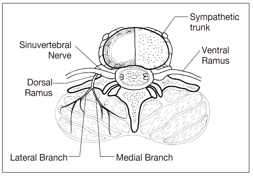
Figure 2
Schematic drawing of sacroiliac joint demonstrating the innervating nerves.
IOL: interosseous ligament, SLB: sacral lateral branch, SN: sacral nerve, SIJ: sacroiliac joint
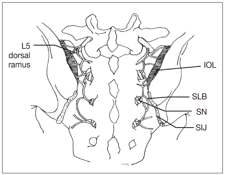
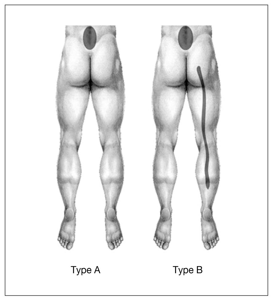
Figure 3
Pain distribution patterns in lumbar zygapophyseal joint dysfunction. Type E (undetermined type) is not depicted here.
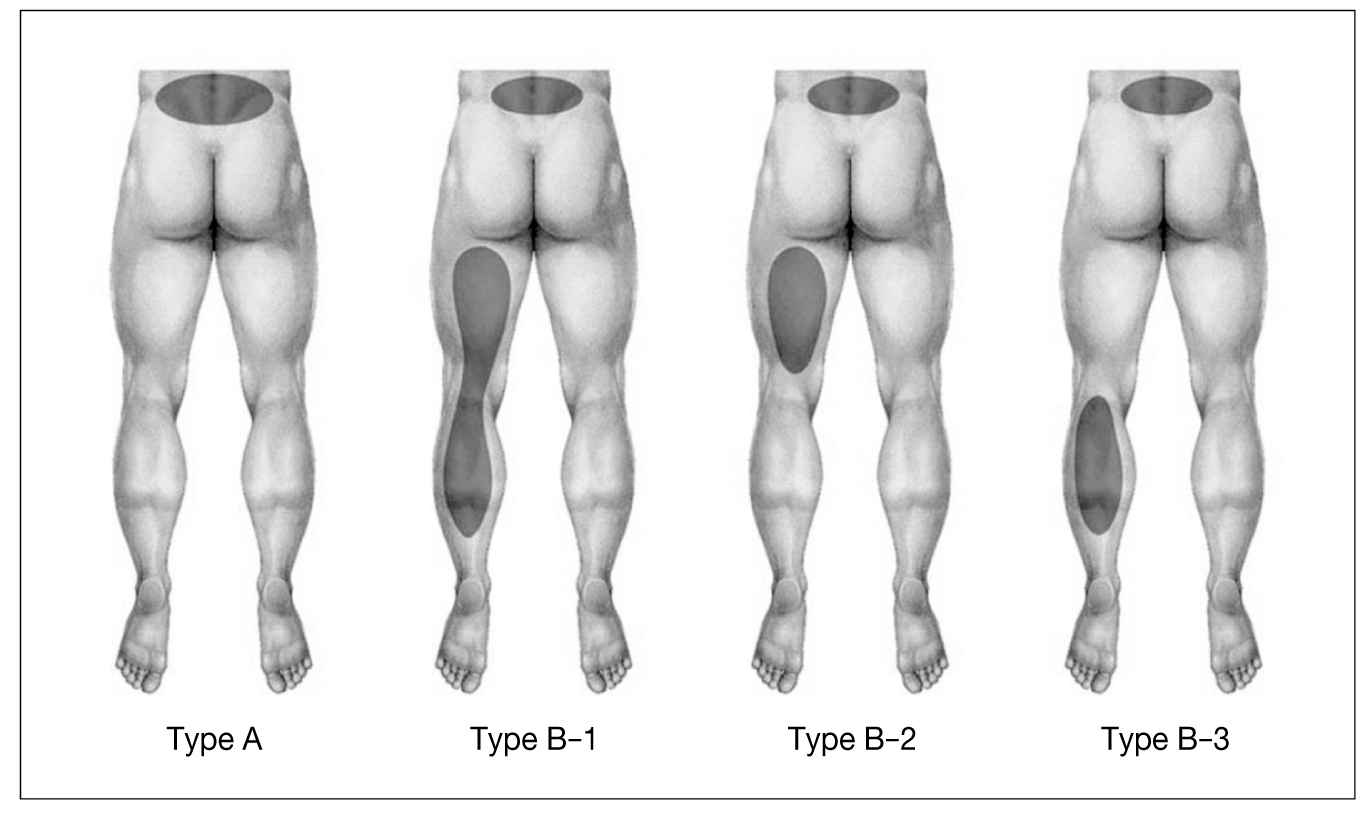
Figure 4
Pain distribution patterns in sacroiliac joint dysfunction. Type E (undetermined type) is not depicted here.
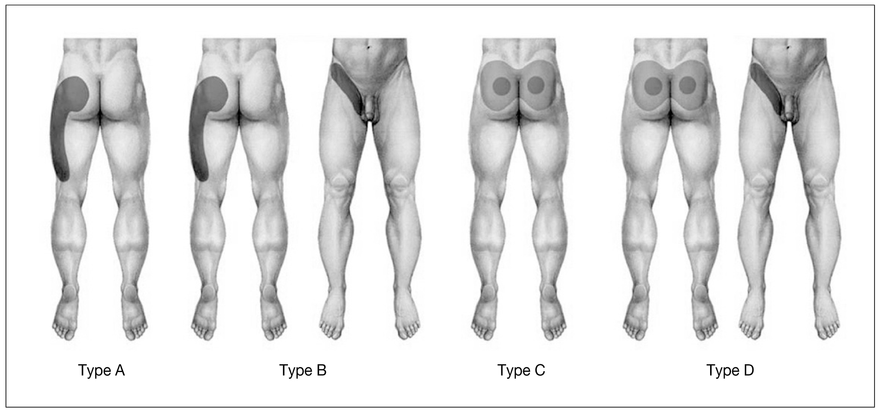
Figure 6
Modified Dallas discogram classification. Note that disk degeneration is confined inside the disk in Grade 1, 2, and 3. Disk herniations are demonstrated in Grade 4 and 5.

Figure 7
Computer screen of automated discography device demonstrating the increase of intradiscal pressure (A) with change of pain
severity (B).
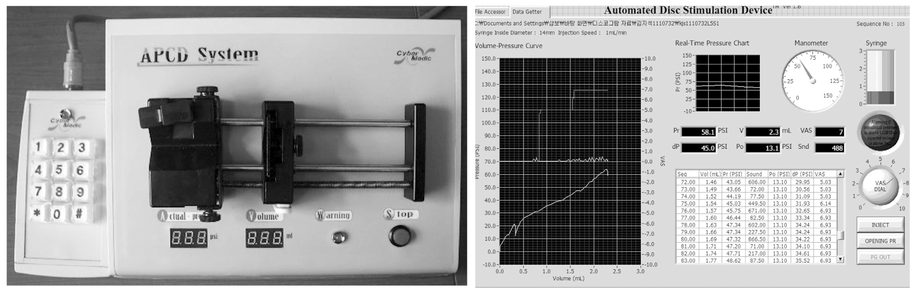
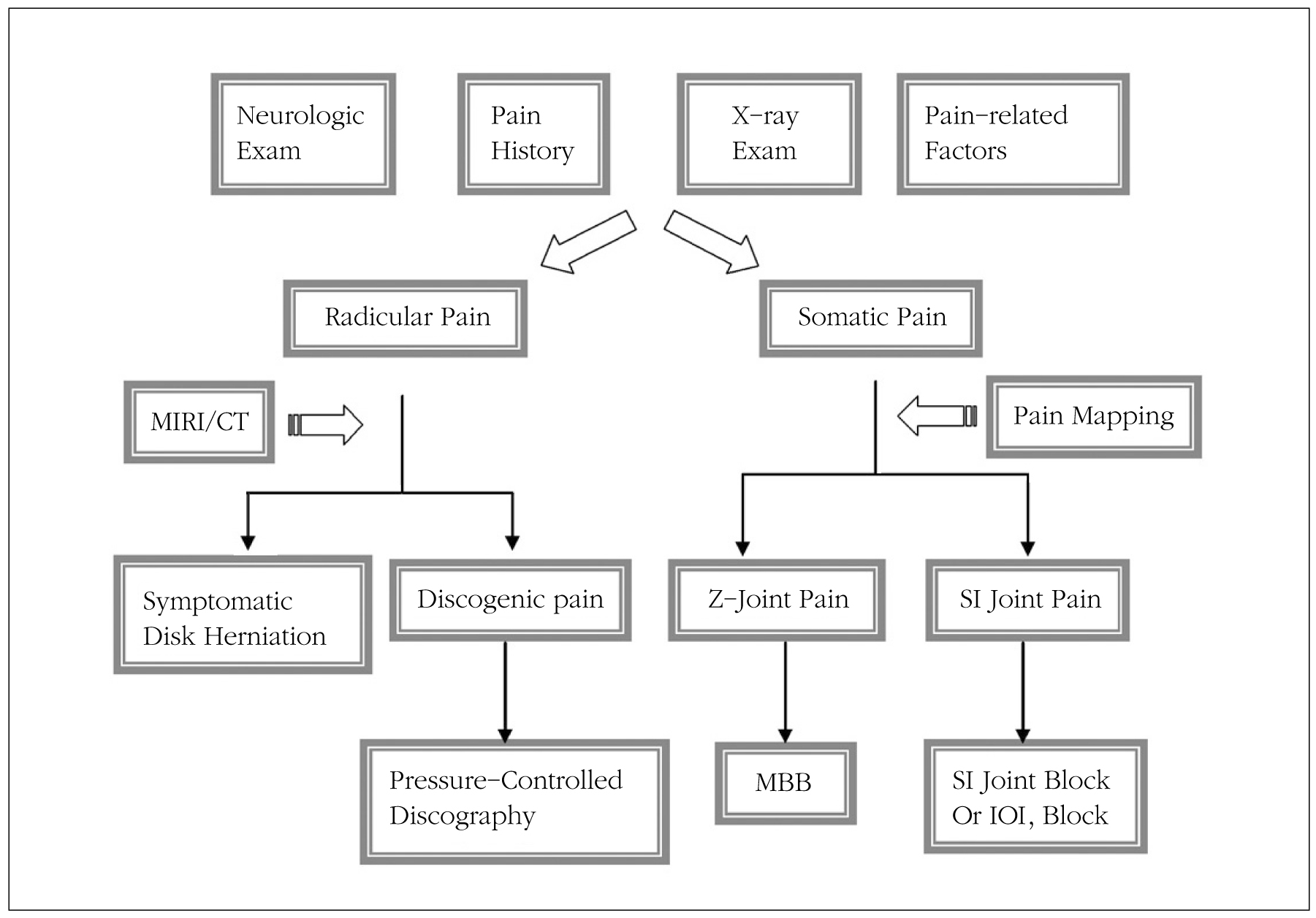
Figure 8
Algorithm for the management of chronic low back pain.
Z-joint: zygapophyseal joint, Exam: examination, SI: sacroiliac, MBB: medial branch block, IOL: interosseous ligament
- TOOLS
-
- Share :
-


-
METRICS

-
- 3 Crossref
- Scopus
- 1,208 View
- 21 Download
-

-
Related articles in
J Korean Med Assoc -
Diagnosis and Treatment of Chronic Congestive Heart Failure1997 November;40(11)
Clinical History and Diagnosis of Chronic Obstructive Pulmonary Disease2006 April;49(4)
Analgesics for Lower Back Pain2006 August;49(8)
- Editorial Office
-
37 Ichon-ro 46-gil, Yongsan-gu, Seoul
Tel: +82-2-6350-6562 Fax: +82-2-792-5208 E-mail: jkmamaster@gmail.com
Copyright © 2024 by Korean Medical Association.
